

Category: Foot Care
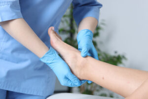
What is a Vascular Test?
A vascular test — also called a circulation test — is a simple, non-invasive screening performed right in the office. Using pressure cuffs and small sensors, it checks blood flow in your arms and legs to catch poor circulation and Peripheral Artery Disease (PAD) early, before it limits your life.
What is PAD?
Peripheral Artery Disease (PAD) happens when the arteries that carry blood to your legs and feet become narrowed or blocked — usually from plaque buildup, the same process that affects the heart. That means less oxygen-rich blood is reaching your limbs. PAD is a progressive, chronic condition, and symptoms often don’t show up until it’s already advanced — which is exactly why early screening matters.
Do Any of These Sound Familiar?
- Cramping, tingling, burning, or heaviness in your feet — while walking or at rest
- Pain when walking or standing
- Cold or numb toes
- Slow-healing wounds
- Swelling in the lower legs
- Skin discoloration
- Unusual hair loss on the feet or legs
Risk Factors to Watch For
- History of diabetes
- Smoking, past or present
- Heart disease
- High blood pressure or high cholesterol
- Family history of circulation problems
- Age 50 or older
If any of these apply to you, you could have an underlying circulation problem in your legs and feet — even without obvious symptoms.
Why Circulation Testing Matters
Think of your circulation like the plumbing in your home. Blockages are often subtle and easy to miss — until they cause a sudden, serious problem. The same thing can happen when circulation to your legs, feet, and toes becomes blocked. Left unchecked, it can lead to major complications. Caught early, a simple test can be limb-saving and even lifesaving.
What to Expect
Vascular testing is quick, painless, and performed right in the office — no needles, no downtime. We make it easy, with same-day screening appointments available.
Know Your Results, Know Your Options
Your results help identify the right treatment to get you back to doing the things you enjoy. Ask your foot and ankle doctor about vascular testing, or find out in 2 minutes whether a screening is right for you.
A plantar fibroma is a harmless growth in the arch of the foot, developing within the plantar fascia tissue. It’s typically small, less than 1 inch, and grows slowly over time. While painless in early stages, it may cause discomfort if it enlarges or is pressed. It’s more common in young or middle-aged adults, with a suspected genetic component. Plantar fibroma can progress to plantar fibromatosis, associated with conditions like liver disease and diabetes. Diagnosis involves a physical exam and possibly imaging tests like X-ray or MRI to confirm and rule out other conditions. In rare cases, a biopsy may be performed for further evaluation.
Platelet-rich plasma (PRP) is a treatment where a person’s own blood is used to heal bone or soft tissue injuries. It’s often considered for chronic injuries that have not responded to conservative care, for example Achilles Tendonitis. PRP involves taking a blood sample, spinning it in a centrifuge to isolate platelets, then injecting them into the injured area. After PRP, patients may need to avoid strenuous activity for a short time and may experience temporary increased pain. While more studies are needed, PRP is generally considered low-risk and may improve healing.
Ankle pain can occur suddenly without an obvious injury and may be caused by various conditions such as osteoarthritis, rheumatoid arthritis, reactive arthritis, lupus, gout, bursitis, scleroderma, fallen arches, Achilles tendonitis, osteochondral lesion of the talus, or infections. Treatment depends on the underlying cause and may include medications, physical therapy, orthotics, or surgery. It’s important to seek medical attention for unexplained ankle pain to prevent potential complications.
Heel pain can be caused by various factors, including faulty biomechanics, injury, or wearing poorly constructed footwear. Common causes include heel spurs, which are bony growths under the heel bone, and plantar fasciitis, an inflammation of the connective tissue on the bottom of the foot. Excessive pronation, or inward motion of the foot, can also contribute to heel pain. Achilles tendinitis, rheumatoid arthritis, bursitis, neuroma, Haglund’s deformity, and bone bruises are other possible causes. If heel pain persists, limits daily activities, or is accompanied by inflammation, it’s advisable to consult a podiatrist for diagnosis and treatment.
Plantar fasciitis is a common cause of heel pain affecting around 2 million people each year. It happens when the tissue supporting the arch of your foot gets irritated and inflamed. This tissue, called the plantar fascia, connects your heel to the front of your foot.
The main causes of plantar fasciitis are excessive pressure and strain on the feet, often from activities like walking, running, or playing sports. Factors that can increase your risk include starting new activities, doing repetitive high-impact exercises, standing for long periods on hard surfaces, having flat feet or high arches, tight calf muscles, being overweight, and aging (typically between 40 to 60 years old).
Common symptoms of plantar fasciitis include heel pain, especially in the morning or after long periods of rest, which usually eases after walking a bit. Pain often increases after exercise rather than during it.
When you see a doctor for plantar fasciitis, they’ll check for signs like a high arch or flat foot, tenderness on the bottom of your foot near the heel, and limited upward movement in your ankle. They’ll also rule out other possible causes of heel pain, like Achilles tendonitis or stress fractures in the heel bone.
Regenerative treatments aim to stimulate the body to regenerate or replace cells and tissues damaged due to aging, disease, or injury instead of treating orthopedic conditions through medication and invasive surgical procedures. Such treatments include growth factors, cytokines, collagen, platelet-rich plasma (PRP), amniotic-derived materials, and cell therapy.
Because these are fast and simple treatments, they are a great alternative to surgery for certain conditions. Regenerative treatments may be a good option for foot and ankle conditions such as arthritis, bursitis, diabetic foot ulcers, venous stasis ulcers, fat pad atrophy and associated metatarsalgia, fibroma, joint pain, metatarsalgia, neuroma, nonunions, plantar fasciitis, plantar plate injury, porokeratosis, tendonitis of the foot and ankle, and wounds (chronic and post-operative).
Kinesiology tape is a therapeutic tape that’s applied strategically to the feet and ankles to provide support, lessen pain, reduce swelling, and improve performance. Kinesio tape is made with a proprietary blend of cotton and nylon. It’s designed to mimic the skin’s elasticity so you can use your full range of motion. The tape’s medical-grade adhesive is also water-resistant and strong enough to stay on for three to five days, even while you work out or take showers.
When the tape is applied to your body, it recoils slightly, gently lifting your skin. It is believed that this helps to create a microscopic space between your skin and the tissues underneath it.
Kinesiology tape is used for treating injuries, supporting weak zones of the muscles or joints, re-educating muscles, and enhancing athletic performance.
This test analyzes how you walk and identifies any abnormal walking patterns. A gait analysis has several components. Together they provide a comprehensive picture of the various factors contributing to the gait disorder.
First, the clinical exam is conducted by the doctor to determine the degree of strength, range of motion, tone, and selective motor control in the lower extremity. Then, you will be videotaped while walking. We will also capture video of your standing posture and footwear to assess wear patterns. This is put into a 3-D picture of the kinematics, or joint motion, to see how the body moves while walking.
Reflective markers (small balls) are taped onto the legs, pelvis, and trunk (shoulders and hips). Digital cameras record the markers’ movements to measure joint motion at the trunk, pelvis, hip, knee, and ankle. Data of the forces that cross the joints, kinetics, further reveal factors contributing to abnormal movement.
Finally, EMG provides information about how the muscles work while walking. We use the EMG data to see how each muscle functions and how it contributes to the gait abnormality. Pressure-sensitive foot switches tell us when and what part of the foot contacts the ground while walking and allow the EMG data to be correlated with the stance and swing phases of gait. In total, the gait analysis takes about two hours.